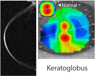
However when those fail to provide adequate correction due to significant astigmatism or are not tolerated anymore, surgery can be considered. Reduced visual acuity can be initially compensated by contact lenses such as rigid gas permeable or scleral contact lenses. Modern topographical measurement devices such as Scheimpflug imaging allow us to distinguish now crab-claw patterns in PMD-like keratoconus from rarer true PMD disease. 1 Some cases of PMD can overlap keratoconus and typical patterns such as “crab-claws” or “kissing birds” patterns can be seen in both diseases as initially described. PMD is distinctive in its features from keratoconus in the way that there is a crescent-shaped inferior band of corneal thinning with associated ectasia and the disease is usually diagnosed in an older set of patients with a male predilection. The disease progression can induce irregular astigmatism leading sometimes to reduced visual acuity. Pellucid marginal degeneration (PMD) is a rare non-inflammatory corneal ectatic disorder that is characterized by progressive peripheral corneal thinning. Keywords: cornea, ectasia, pellucid marginal degeneration, cornea cross-linking, wedge resection Long-term follow-up shows stabilization and absence of regression in the second eye up to eight months after the surgery.Ĭonclusion: Combining corneal cross-linking with corneal wedge resection in the case of advanced pellucid marginal degeneration patients could be a good option in order to stabilize topographical and refractive results and reduces the risk of regression. The second eye is then treated with the same surgical technique combined with cornea cross-linking. The first eye shows initial good results however after few months regression is observed. In order to avoid using donated tissue through corneal grafting we decide to perform a sectorial lamellar crescentric wedge excision of the thinner inferior part of the cornea involving the pellucid marginal degeneration and suture it. As he is now intolerant to contact lenses a surgical option is offered to him.

We present here the use of corneal cross-linking in order to stabilize the parameters on the long term.Ĭase presentation: We present here the case of a 53 years old patient with bilateral advanced pellucid marginal degeneration. However topographical and refractive results are in some instance fluctuating. Corneal wedge resection allows for good visual rehabilitation without the risks of tissue rejection. It is traditionally managed with corneal transplantation, however alternative surgical options exist. Ophthalmology Department, University of Lausanne, Jules Gonin Eye Hospital, Lausanne, SwitzerlandĬorrespondence: George Kymionis Hôpital Ophtalmique Jules Gonin, 15 avenue de France, Lausanne 1004 Switzerlandīackground: Advanced pellucid marginal degeneration is a debilitating disease that warrants the use of surgery when the visual acuity is reduced and contact lenses are not tolerated anymore. George Kymionis, Nafsika Voulgari, Erwin Samutelela, George Kontadakis, David Tabibian


 0 kommentar(er)
